KC-1456

293T-ISRE-Luc2-ADAR1-P150 Cell Line

×
22175
Home » Cell Lines » 293T-ISRE-Luc2-ADAR1-P150 Cell Line

Background of 293T-ISRE-Luc2-ADAR1-P150 Cell Line

ADAR (adenosine deaminase RNA specific; important alias: ADAR1) encodes adenosine deaminase acting on RNA 1, an interferon-inducible enzyme that catalyzes adenosine-to-inosine (A-to-I) RNA editing on double-stranded RNA substrates. The p150 isoform is the full-length, interferon-inducible form that localizes to both cytoplasm and nucleus. ADAR1 plays critical roles in innate immune regulation, preventing aberrant activation of the interferon response by self RNA, and is implicated in cancer progression and autoimmune diseases such as Aicardi-Goutières syndrome. It represents an emerging therapeutic target in oncology and immunology.

Specifications

Catalog NumberKC-1456
Cell Line Name293T-ISRE-Luc2-ADAR1-P150 Cell Line
NCBI/UniProt Accession NumberNM_001111.5
Host Cell Line293T
DescriptionHEK293T reporter cell line stably expressing ISRE-driven Luc2 reporter and human ADAR1-P150, designed for interferon pathway and ADAR1 functional studies
QuantityOne vial of frozen cells (≥2×10⁶ cells)
StabilityStable in culture over a minimum of 10 passages
ApplicationDrug screening and biological assays
Freezing Medium70% DMEM+20% FBS+10% DMSO
Propagation MediumDMEM+10%FBS+140μg/mL Hygromycin B
Selection MarkerHygromycin B
MorphologyEpithelial-like
SubcultureSplit at a ratio of 1:5–1:8 every 2–3 days
Incubation37°C with 5% CO₂
StorageLiquid nitrogen immediately upon receiving
Doubling TimeEstimated 16–31 hours
Mycoplasma StatusNegative
In Vivo ValidationNA

Cell Line Generation

The 293T-ISRE-Luc2-ADAR1-P150 cell line was generated using a lentiviral vector expressing the ISRE-Luc2 reporter and human ADAR1-P150 sequences.

Characterization

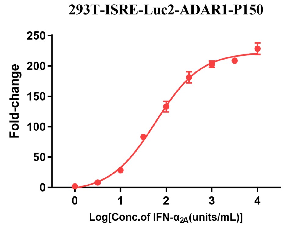
Figure 1: 293T-ISRE-Luc2-ADAR1-P150 cells were seeded into the 96-well plate, and treated with the highest concentration 10000 units/mL Interferon Alpha 2A, 3.16 times diluted to 9 concentrations. After reaction for 16 hours, readout with Bright-lite Luciferase Assay system.

Cell Resuscitation

  1. Pre-warm complete culture medium (basal medium and 10% FBS) in a 37°C water bath.
  2. Rapidly thaw the cryovial in a 37°C water bath for 1-2 minutes with gentle agitation.
  3. Transfer the vial to a biosafety cabinet, and disinfect the exterior with 70% ethanol.
  4. Aseptically transfer the cell suspension dropwise into a sterile centrifuge tube containing 9.0 mL of pre-warmed complete medium.
  5. Centrifuge at approximately 125 × g for 5–7 minutes at room temperature, carefully aspirate the supernatant without disturbing the cell pellet.
  6. Gently resuspend the pellet in an appropriate volume of complete medium and transfer the suspension into a T25 flask.
  7. Incubate the flask in a 37°C in a humidified 5% CO₂ incubator.
  8. Assess cell viability and morphology after 24 hours. If cells appear healthy, replace the medium with fresh medium supplemented with the appropriate selective antibiotic.
  9. Subculture the cells at a ratio of 1:5–1:8 every 2–3 days upon reaching 80%–90% confluency.

Cell Freezing

  1. Prepare the freezing medium (70% basal medium, 20% FBS and 10% DMSO) freshly before use.
  2. Pre-chill the freezing medium on ice and label the cryovials accordingly.
  3. Transfer the cell suspension to a sterile conical tube and perform a cell count to determine total viability and density.
  4. Centrifuge the cells at 250×g for 5 minutes at room temperature; carefully aspirate the supernatant.
  5. Gently resuspend the pellet in chilled freezing medium, ensuring a minimum cell density of 3×10⁶ cells/mL.
  6. Aliquot 1 mL of the cell suspension into each pre-labeled cryovial.
  7. Place the cryovials into a CoolCell® container and store at -80°C overnight for controlled-rate cooling.
  8. Transfer the cryovials to the liquid nitrogen for long-term storage the following day.

References

  1. Nishikura K. Functions and regulation of RNA editing by ADAR deaminases. Annu Rev Biochem. 2010;79:321-349.
  2. Pfaller CK, et al. ADAR1 editing and its role in cancer. Cancer Cell. 2021;39(2):178-193.
  3. Mannion NM, et al. The RNA editing enzyme ADAR1: more than just an editor. IUBMB Life. 2020;72(4):775-788.

Use License Agreement

Research Use Only.
Not for use in diagnostic procedures or therapeutic applications.
Redistribution of the cell line or its derivatives is prohibited without prior written permission from Kyinno Biotechnology.