KC-3107

MC38-PDL1-CLDN18.2-OS8 Cell Line

×
26895
Home » MC38-PDL1-CLDN18.2-OS8 Cell Line

Background of MC38-PDL1-CLDN18.2-OS8 Cell Line

PD-L1 (Programmed Death-Ligand 1) , also known as CD274 or B7-H1, is a type I transmembrane immune checkpoint ligand encoded on chromosome 9p24.1. It binds PD-1 on T cells to deliver inhibitory signals, suppressing T-cell activation and cytokine production; in tumors, this axis enables immune evasion. PD-L1 is overexpressed in many malignancies, including colon cancer, and its expression is prognostic in several human cancers.
CLDN18.2 (Claudin-18 isoform 2) is a tight junction protein generated by alternative splicing of CLDN18. It is almost exclusively expressed on gastric mucosal tight junctions, with minimal expression in other healthy adult tissues. CLDN18.2 is aberrantly overexpressed in gastric, gastroesophageal junction, pancreatic, and ovarian cancers, functioning as a barrier component and a tumor-associated antigen.
OS8 is an engineered membrane-anchored chimeric T cell engager, constructed by fusing the single-chain variable fragment (scFv) of the anti-human CD3 monoclonal antibody OKT3 to the transmembrane and cytoplasmic domains of CD8α. When displayed on the surface of a cell line, OS8 functions as an artificial antigen-presenting cell component that directly engages and activates the TCR on T cells, enabling T-cell-based functional assays.

Specifications

Catalog NumberKC-3107
Cell Line NameMC38-PDL1-CLDN18.2-OS8 Cell Line
NCBI/UniProt Accession NumberNM_014143.4, NM_001002026
Clone Number5#
Host Cell LineMC38-mouse-PDL1-KO-PDL1
DescriptionStable MC38 cell line expressing exogenous human PDL1 and CLDN18.2 genes, together with a membrane-anchored OS8 T cell engager construct.
QuantityOne vial of frozen cells (≥2×10⁶ cells)
StabilityStable in culture over a minimum of 10 passages
ApplicationDrug screening and biological assays
Freezing MediumDMEM+20% FBS+10% DMSO
Propagation MediumDMEM+10%FBS+5µg/mL Puromycin
Selection MarkerPuromycin
MorphologyEpithelial
SubcultureSplit saturated culture 1:3-1:5 every 2-3 days
Incubation37 °C with 5% CO2
StorageLiquid nitrogen immediately upon receiving
Doubling TimeApproximately 35 hours
Mycoplasma StatusNegative
In Vivo ValidationNA

Cell Line Generation

The MC38-PDL1-CLDN18.2-OS8 cell line was generated by lentiviral transduction of MC38 cells to stably express human PDL1, CLDN18.2, and the OS8 membrane-anchored T cell engager.

Characterization

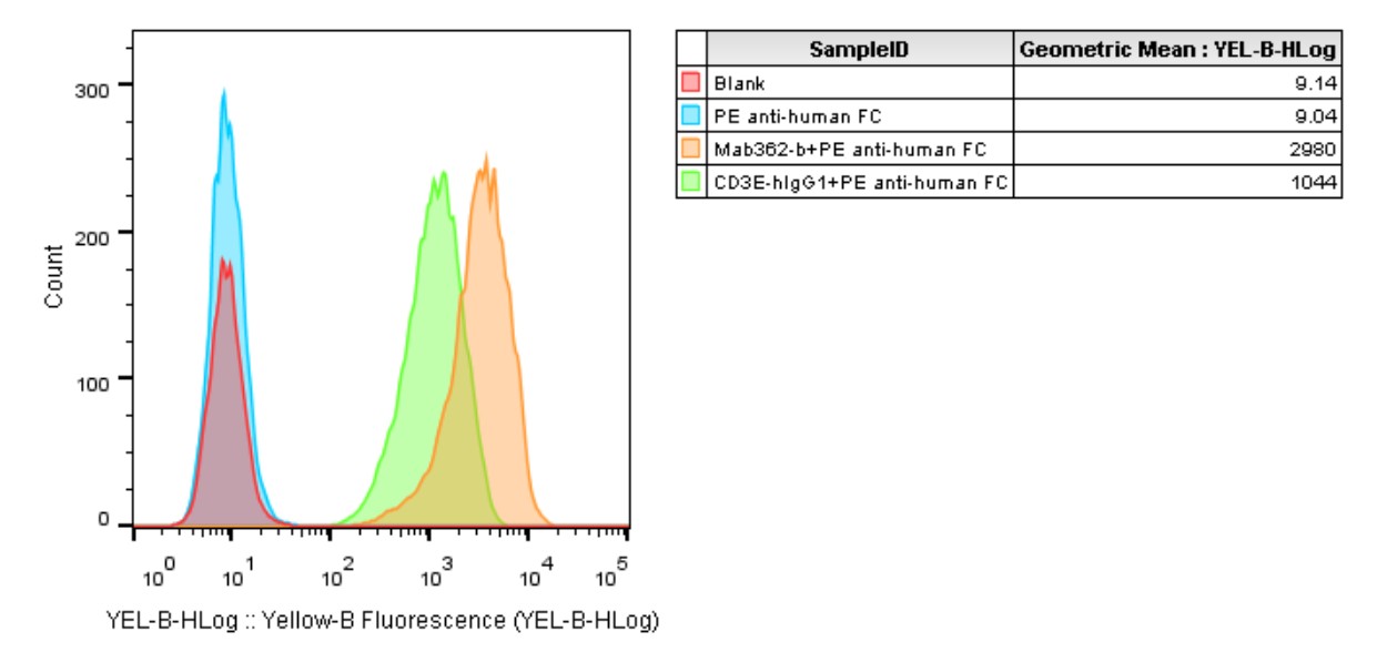
Figure 1: Characterization of CLDN18.2 (orange) and OS8 (green) overexpression in the MC38-PDL1-CLDN18.2-OS8 stable clone using FACS.

Cell Resuscitation

  1. Pre-warm complete medium (basal medium supplemented with 10% FBS and other required supplements, without selective antibiotic) in a 37°C water bath.
  2. Rapidly thaw the cryovial in a 37°C water bath for 1–2 min with gentle agitation.
  3. Transfer the vial into a biosafety cabinet and disinfect the exterior surface with 70% ethanol.
  4. Aseptically transfer the cell suspension dropwise into a sterile centrifuge tube containing 9.0 mL of pre-warmed complete medium.
  5. Centrifuge at approximately 125 × g for 5–7 min at room temperature; carefully aspirate the supernatant without disturbing the cell pellet.
  6. Gently resuspend the pellet in an appropriate volume of pre-warmed complete medium and transfer the suspension into a T25 flask.
  7. Incubate the flask in a humidified 37°C, 5% CO2 incubator.
  8. Assess cell morphology and viability after 24 h. If cells are in good condition, replace the medium with complete fresh medium supplemented with the appropriate selective antibiotic.
  9. Subculture the cells at a split ratio of 1:3–1:5 every 2–3 days once cells reach 80%–90% confluency.

Cell Freezing

  1. Prepare the freezing medium (70% basal medium, 20% FBS and 10% DMSO) freshly before use.
  2. Pre-chill the freezing medium on ice and label the cryovials accordingly.
  3. Transfer the cell suspension to a sterile conical tube and perform a cell count to determine total viability and density.
  4. Centrifuge the cells at 250×g for 5 minutes at room temperature; carefully aspirate the supernatant.
  5. Gently resuspend the cell pellet in chilled freezing medium, ensuring a minimum cell density of 3×10⁶ cells/mL.
  6. Aliquot 1 mL of the cell suspension into each pre-labeled cryovial.
  7. Place the cryovials into a CoolCell® container and store at -80°C overnight for controlled-rate cooling.
  8. Transfer the cryovials to the liquid nitrogen for long-term storage the following day.

References

1. Dominguez Wiscovitch A, Sanchez Mendez RJ, Chuy J. CLDN18.2-Targeted Therapy in Gastrointestinal Cancers. Cancers (Basel). 2025 Nov 25;17(23):3764. doi: 10.3390/cancers17233764. PMID: 41374966; PMCID: PMC12691406.
2. Zhang F, Okazaki U, Nakayama I, Shitara K. CLDN18.2 in Gastric Cancer: Current Therapeutic Landscape and Future Perspectives. J Gastric Cancer. 2026 Jan;26(1):127-145. doi: 10.5230/jgc.2026.26.e10. PMID: 41517852; PMCID: PMC12802060.
3. Shah MA, Kennedy EB, Deighton D, Jhawer M, Kadakia KC, Mukherjee S, Nevala-Plagemann C, Saeed A, Schneider BJ, Semrad T, Shitara K, Tenner L, Uboha NV, Vincelli M, Zohrabyan D, Rajdev L. Immunotherapy and Targeted Therapy for Advanced Gastroesophageal Cancer: ASCO Guideline Update. J Clin Oncol. 2026 Apr 20;44(12):1145-1165. doi: 10.1200/JCO-25-02958. Epub 2026 Feb 26. PMID: 41747202.
4. Lee H, Lee Y, Chung J. Characterization of anti-CD3 antibodies in clinically available bispecific T cell engagers. Semin Hematol. 2025 Aug;62(4):279-288. doi: 10.1053/j.seminhematol.2025.08.004. Epub 2025 Aug 29. PMID: 40987715.

Use License Agreement

Research Use Only.
Not for use in diagnostic procedures or therapeutic applications.
Redistribution of the cell line or its derivatives is prohibited without prior written permission from Kyinno Biotechnology.