KC-4038

293T-CDH16 Cell Line

×
40534
Home » 293T-CDH16 Cell Line

Background of 293T-CDH16 Cell Line

CDH16 encodes cadherin 16. Mapped to a previously identified cluster of cadherin genes on chromosome 16q22.1, the gene localizes with superfamily members CDH1, CDH3, CDH5, CDH8 and CDH11. The protein consists of an extracellular domain containing 6 cadherin domains, a transmembrane region and a truncated cytoplasmic domain but lacks the prosequence and tripeptide HAV adhesion recognition sequence typical of most classical cadherins. Expression is exclusively in kidney, where the protein functions as the principal mediator of homotypic cellular recognition, playing a role in the morphogenic direction of tissue development. Alternatively spliced transcript variants encoding distinct isoforms have been identified.

Specifications

Catalog NumberKC-4038
Cell Line Name293T-CDH16 Cell Line
NCBI/UniProt Accession NumberNM_004062.3
Clone Number4#
Host Cell Line293T
Description293T-CDH16 is a stable engineered cell line expressing human CDH16
QuantityOne vial of frozen cells (≥2×10⁶ cells/vial)
StabilityStable in culture over a minimum of 10 passages
ApplicationDrug screening and biological assays
Freezing Medium70% DMEM+20% FBS+10% DMSO
Propagation MediumDMEM+10%FBS+1μg/mL Puromycin
Selection MarkerPuromycin
MorphologyEpithelial
SubcultureSplit at a ratio of 1:4–1:6 every 2–3 days
Incubation37 °C with 5% CO₂
StorageLiquid nitrogen immediately upon receiving
Doubling TimeEstimated 19–36 hours
Mycoplasma StatusNegative
In Vivo ValidationNA

Cell Line Generation

293T-CDH16 cell line was generated using a lentiviral vector expressing the human CDH16 sequence.

Characterization

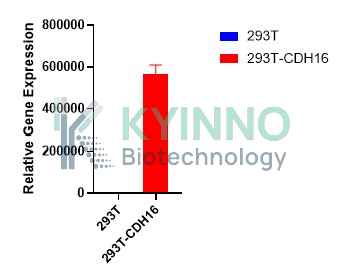
Figure 1: Characterization of human CDH16 overexpression in the 293T-CDH16 stable clone using qPCR.

Figure 2: Characterization of human CDH16 overexpression in the 293T-CDH16 stable clone using PCR sequence.

Cell Resuscitation

  1. Pre-warm complete culture medium (basal medium and 10% FBS) in a 37°C water bath.
  2. Rapidly thaw the cryovial in a 37°C water bath for 1-2 minutes with gentle agitation.
  3. Transfer the vial to a biosafety cabinet, and disinfect the exterior with 70% ethanol.
  4. Aseptically transfer the cell suspension dropwise into a sterile centrifuge tube containing 9.0 mL of pre-warmed complete medium.
  5. Centrifuge at approximately 125 × g for 5–7 minutes at room temperature, carefully aspirate the supernatant without disturbing the cell pellet.
  6. Gently resuspend the pellet in an appropriate volume of complete medium and transfer the suspension into a T25 flask.
  7. Incubate the flask in a 37°C in a humidified 5% CO₂ incubator.
  8. Assess cell viability and morphology after 24 hours. If cells appear healthy, replace the medium with fresh medium supplemented with the appropriate selective antibiotic.
  9. Subculture the cells at a ratio of 1:4–1:6 every 2–3 days upon reaching 80%–90% confluency.

Cell Freezing

  1. Prepare the freezing medium (70% basal medium, 20% FBS and 10% DMSO) freshly before use.
  2. Pre-chill the freezing medium on ice and label the cryovials accordingly.
  3. Transfer the cell suspension to a sterile conical tube and perform a cell count to determine total viability and density.
  4. Centrifuge the cells at 250×g for 5 minutes at room temperature; carefully aspirate the supernatant.
  5. Gently resuspend the pellet in chilled freezing medium, ensuring a minimum cell density of 3×10⁶ cells/mL.
  6. Aliquot 1 mL of the cell suspension into each pre-labeled cryovial.
  7. Place the cryovials into a CoolCell® container and store at -80°C overnight for controlled-rate cooling.
  8. Transfer the cryovials to the liquid nitrogen for long-term storage the following day.

References

  1. Siraj AK, Parvathareddy SK, Al-Rasheed M et al. Loss of CDH16 expression is a strong independent predictor for lymph node metastasis in Middle Eastern papillary thyroid cancer. Scientific reports. 2023 Oct 29;13(1):18559. PMID: 37899424. doi: 10.1038/s41598-023-45882-x.
  2. Lennartz M, Csomós H, Chirico V et al. Cadherin-16 (CDH16) immunohistochemistry: a useful diagnostic tool for renal cell carcinoma and papillary carcinomas of the thyroid. Scientific reports. 2023 Aug 9;13(1):12917. PMID: 37558687. doi: 10.1038/s41598-023-39945-2.
  3. Lennartz M, Csomós H, Bernreuther C et al. Reduced CDH16 expression is linked to poor prognosis in clear cell renal cell carcinoma 16. Urologic oncology. 2022 Jul;40(7):348.e1-348.e8. PMID: 35606285. doi: 10.1016/j.urolonc.2022.03.017.

Use License Agreement

Research Use Only.
Not for use in diagnostic procedures or therapeutic applications.
Redistribution of the cell line or its derivatives is prohibited without prior written permission from Kyinno Biotechnology.