KC-3924

293T-mouse-TEK Cell Line

×
40316
Home » 293T-mouse-TEK Cell Line

Background of 293T-mouse-TEK Cell Line

Tyrosine-protein kinase that acts as cell-surface receptor for ANGPT1, ANGPT2 and ANGPT4 and regulates angiogenesis, endothelial cell survival, proliferation, migration, adhesion and cell spreading, reorganization of the actin cytoskeleton, but also maintenance of vascular quiescence. Has anti-inflammatory effects by preventing the leakage of pro-inflammatory plasma proteins and leukocytes from blood vessels. Required for normal angiogenesis and heart development during embryogenesis.

Specifications

Catalog NumberKC-3924
Cell Line Name293T-mouse-TEK Cell Line
NCBI/UniProt Accession NumberNM_001290549.1
Clone Number1#
Host Cell Line293T
DescriptionStable 293T cell line expressing exogenous mouse TEK gene
QuantityOne vial of frozen cells (≥2×10⁶ cells)
StabilityStable in culture over a minimum of 10 passages
ApplicationDrug screening and biological assays
Freezing Medium70% basal medium+20% FBS+10% DMSO
Propagation MediumDMEM+10% FBS +1μg/mL Puromycin
Selection MarkerPuromycin
MorphologyEpithelial-like
SubcultureSplit saturated culture 1:4-1:8 every 2-3 days
Incubation37 °C with 5% CO2
StorageLiquid nitrogen immediately upon receiving
Doubling TimeApproximately 30 hours
Mycoplasma StatusNegative
In Vivo ValidationNA

Cell Line Generation

293T-mouse-TEK cell line was generated using a lentiviral vector expressing the mouse TEK sequence.

Characterization

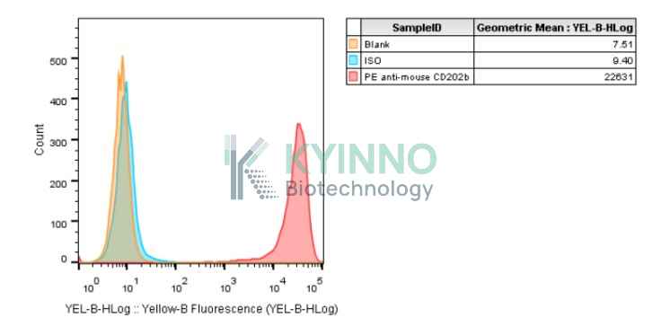
Figure 1: Characterization of mouse TEK overexpression in the 293T-mouse-TEK stable clone using FACS.

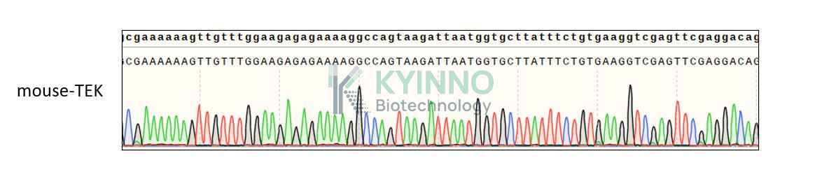
Figure 2: Characterization of mouse TEK in the 293T-mouse-TEK stable clone using PCR sequencing.

Cell Resuscitation

  1. Pre-warm complete culture medium (DMEM+10% FBS +1μg/mL Puromycin) in a 37°C water bath.
  2. Rapidly thaw the cryovial in a 37°C water bath for 1-2 minutes with gentle agitation.
  3. Transfer the vial to a biosafety cabinet, and disinfect the exterior with 70% ethanol.
  4. Aseptically transfer the cell suspension dropwise into a sterile centrifuge tube containing 9.0 mL of pre-warmed complete medium.
  5. Centrifuge at approximately 125 × g for 5–7 minutes at room temperature, carefully aspirate the supernatant without disturbing the cell pellet.
  6. Gently resuspend the pellet in an appropriate volume of complete medium and transfer the suspension into a T25 flask.
  7. Incubate the flask in a 37°C in a humidified 5% CO2 incubator.
  8. Assess cell viability and morphology after 24 hours. If cells appear healthy, replace the medium with fresh medium supplemented with the appropriate selective antibiotic.
  9. Subculture the cells at a ratio of 1:4-1:8 every 2-3 days upon reaching 80%–90% confluency.

Cell Freezing

  1. Prepare the freezing medium (70% basal medium, 20% FBS and 10% DMSO) freshly before use.
  2. Pre-chill the freezing medium on ice and label the cryovials accordingly.
  3. Transfer the cell suspension to a sterile conical tube and perform a cell count to determine total viability and density.
  4. Centrifuge the cells at 250×g for 5 minutes at room temperature; carefully aspirate the supernatant.
  5. Gently resuspend the cell pellet in chilled freezing medium, ensuring a minimum cell density of 3×10⁶ cells/mL.
  6. Aliquot 1 mL of the cell suspension into each pre-labeled cryovial.
  7. Place the cryovials into a CoolCell® container and store at -80°C overnight for controlled-rate cooling.
  8. Transfer the cryovials to the liquid nitrogen for long-term storage the following day.

References

1. Rajasundaram S, Zebardast N, Mehta P, Khawaja AP, Warwick A, Duchinski K, Burgess S, Gill D, Segrè AV, Wiggs J. TIE1 and TEK signalling, intraocular pressure, and primary open-angle glaucoma: a Mendelian randomization study. J Transl Med. 2023 Nov 24;21(1):847.
2. Ha M, Son YR, Kim J, Park SM, Hong CM, Choi D, Kang W, Kim JH, Lee KJ, Park D, Han ME, Oh SO, Lee D, Kim YH. TEK is a novel prognostic marker for clear cell renal cell carcinoma. Eur Rev Med Pharmacol Sci. 2019 Feb;23(4):1451-1458.
3. Mäe MA, He L, Nordling S, Vazquez-Liebanas E, Nahar K, Jung B, Li X, Tan BC, Chin Foo J, Cazenave-Gassiot A, Wenk MR, Zarb Y, Lavina B, Quaggin SE, Jeansson M, Gu C, Silver DL, Vanlandewijck M, Butcher EC, Keller A, Betsholtz C. Single-Cell Analysis of Blood-Brain Barrier Response to Pericyte Loss. Circ Res. 2021 Feb 19;128(4):e46-e62.

Use License Agreement

Research Use Only.
Not for use in diagnostic procedures or therapeutic applications.
Redistribution of the cell line or its derivatives is prohibited without prior written permission from Kyinno Biotechnology.