KC-0889

HCT15-PDL1-uPA Cell Line

×
21189
Home » HCT15-PDL1-uPA Cell Line

Background of HCT15-PDL1-uPA Cell Line

PD-L1, also called human programed cell death ligand 1, is a transmembrane protein that play a major role in suppressing the immune system during particular events such as pregnancy, tissue allografts, autoimmune disease, virus infection and cancer. PD-L1 binds to its receptor PD-1 on activated T cells, B cells, and myeloid cells, to modulate activation or inhibition. Upregulation of PD-L1 can allow cancer cell to evade the host immune system. Urokinase, also known as urokinase-type plasminogen activator (uPA), is a serine protease in blood and urine. Elevated expression of urokinase was found to be correlated with tumor malignancy, and used as a diagnostic biomarker in breast cancer.

Specifications

Catalog NumberKC-0889
Cell Line NameHCT15-PDL1-uPA Cell Line
NCBI/UniProt Accession NumberNM_014143 & NM_002658
Clone Number2#
Host Cell LineHCT15
DescriptionHCT15 cell line stably expressing exogenous PDL1 and uPA gene
QuantityOne vial of frozen cells (≥2×10⁶ cells)
StabilityStable in culture over a minimum of 10 passages
ApplicationDrug screening and biological assays
Freezing Medium70% RPMI1640 + 20% FBS + 10% DMSO
Propagation MediumRPMI1640+10%FBS+100µg/mL Hygromycin B
Selection MarkerHygromycin B
MorphologyEpithelial
SubcultureSplit saturated culture 1:4-1:5 every 2-3 days; seed out at about 1-3 × 10⁵ cells/mL
Incubation37 °C with 5% CO2
StorageLiquid nitrogen immediately upon receiving
Doubling TimeApproximately 30 hours
Mycoplasma StatusNegative
In Vivo ValidationNA

Cell Line Generation

HCT15-PDL1-uPA cell line was generated using a lentiviral vector expressing the PDL1 and uPA sequence.

Characterization

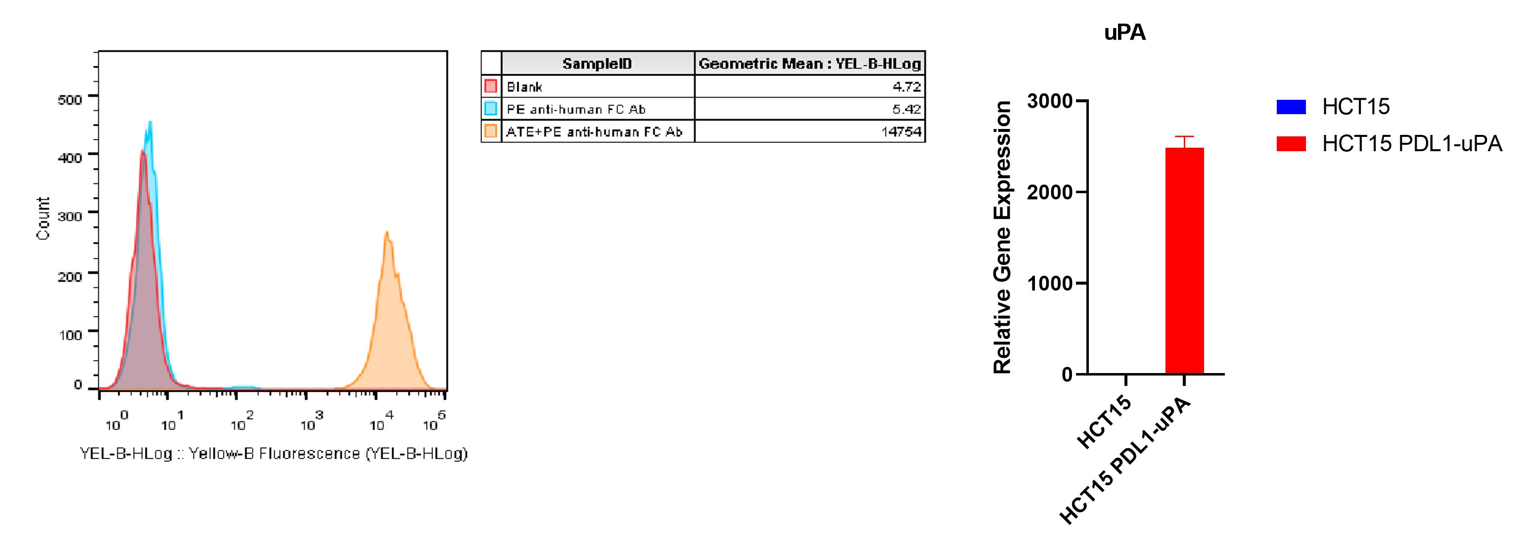
Figure1: Characterization of PDL1 and uPA overexpressing in HCT15 stable clones using FACS and qPCR.

Cell Resuscitation

  1. Prewarm culture medium (RPMI1640+10%FBS+100µg/mL Hygromycin B) in a 37°C water bath.
  2. Thaw the frozen vial in a 37°C water bath for 1-2 minutes.
  3. Transfer the vial into biosafety cabinet, and wipe the surface with 70% ethanol.
  4. Unscrew the top of the vial and transfer the cell suspension gently into a sterile centrifuge tube containing 9.0mL complete culture medium.
  5. Spin at ~ 125 × g for 5-7 minutes at room temperature, and discard the supernatant without disturbing the pellet.
  6. Resuspend cell pellet with the appropriate volume of complete medium and transfer the cell suspension into a T25 culture flask.
  7. Incubate the flask at 37°C, 5% CO2 incubator.
  8. Split saturated culture 1:4-1:5 every 2-3 days; seed out at about 1-3 × 10⁵ cells/mL.

Cell Freezing

  1. Prepare the freezing medium (70% DMEM + 20% FBS + 10% DMSO) fresh immediately before use.
  2. Keep the freezing medium on ice and label cryovials.
  3. Transfer cells to a sterile, conical centrifuge tube, and count the cells.
  4. Centrifuge the cells at 250×g for 5 minutes at room temperature and carefully aspirate off the medium.
  5. Resuspend the cells at a density of at least 3×10⁶ cells/mL in chilled freezing medium.
  6. Aliquot 1 mL of the cell suspension into each cryovial.
  7. Freeze cells in the CoolCell freezing container overnight in a -80°C freezer.
  8. Transfer vials to liquid nitrogen for long-term storage.

References

  1. Sunshine, J. & Taube, J. M. PD-1/PD-L1 inhibitors. Current Opinion in Pharmacology 23, 32–38 (2015).
  2. Boussiotis, V. A. Molecular and Biochemical Aspects of the PD-1 Checkpoint Pathway. N Engl J Med 375, 1767–1778 (2016).
  3. Topalian, S. L. et al. Safety, activity, and immune correlates of anti-PD-1 antibody in cancer. N Engl J Med 366, 2443–2454 (2012).

Use License Agreement

Research Use Only.
Not for use in diagnostic procedures or therapeutic applications.
Redistribution of the cell line or its derivatives is prohibited without prior written permission from Kyinno Biotechnology.