KC-1316

CHOK1-CD40L-Cell-Line

×
21913
Home » CHOK1-CD40L-Cell-Line

Background of CHOK1-CD40L-Cell-Line

CD40L (CD40 Ligand), also known as CD154, TNFSF5, TRAP, or gp39, is a type II membrane glycoprotein of the TNF superfamily, encoded on chromosome Xq26.3. It exists in both membrane-bound and soluble forms. CD40L is predominantly and transiently expressed on activated CD4+ T cells and platelets during immune responses, with lower expression on NK cells, CD8+ T cells, and other leukocytes. It binds CD40 on antigen-presenting cells, activating NF-κB and MAPK pathways to drive B cell activation, antibody class switching, and macrophage differentiation. CD40L is a critical immune checkpoint and therapeutic target. Agonistic anti-CD40 antibodies (e.g., selicrelumab) are in immuno-oncology trials, while antagonistic agents (e.g., dapirolizumab, frexalimab) are being developed for autoimmune diseases such as lupus and multiple sclerosis.

Specifications

Catalog NumberKC-1316
Cell Line NameCHOK1-CD40L-Cell-Line
NCBI/UniProt Accession NumberNM_000074.3
Clone Number4#
Host Cell LineChinese hamster ovary CHO-K1 cell line
DescriptionStable CHOK1 cell line expressing exogenous human CD40L gene
QuantityOne vial of frozen cells (≥2×10⁶ cells)
StabilityStable in culture over a minimum of 10 passages
ApplicationDrug screening and biological assays
Freezing Medium70% RPMI1640+20% FBS+10% DMSO
Propagation MediumRPMI1640+10%FBS+10µg/mL Puromycin
Selection MarkerPuromycin
MorphologyEpithelial-like
SubcultureSplit saturated culture 1:4-1:8 every 2-3 days
Incubation37 °C with 5% CO2
StorageLiquid nitrogen immediately upon receiving
Doubling TimeApproximately 24 hours
Mycoplasma StatusNegative
In Vivo ValidationNA

Cell Line Generation

The CHOK1-CD40L cell line was generated using a lentiviral vector expressing the human CD40L sequence.

Characterization

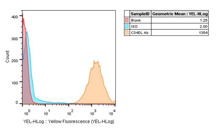
Figure 1: Characterization of CD40L overexpression in the CHOK1-CD40L stable clone using FACS.

Cell Resuscitation

  1. Pre-warm complete culture medium (basal medium and 10% FBS) in a 37°C water bath.
  2. Rapidly thaw the cryovial in a 37°C water bath for 1-2 minutes with gentle agitation.
  3. Transfer the vial to a biosafety cabinet, and disinfect the exterior with 70% ethanol.
  4. Aseptically transfer the cell suspension dropwise into a sterile centrifuge tube containing 9.0 mL of pre-warmed complete medium.
  5. Centrifuge at approximately 125 × g for 5–7 minutes at room temperature, carefully aspirate the supernatant without disturbing the cell pellet.
  6. Gently resuspend the pellet in an appropriate volume of complete medium and transfer the suspension into a T25 flask.
  7. Incubate the flask in a 37°C in a humidified 5% CO2 incubator.
  8. Assess cell viability and morphology after 24 hours. If cells appear healthy, replace the medium with fresh medium supplemented with the appropriate selective antibiotic.
  9. Subculture the cells at a ratio of 1:4-1:8 every 2-3 days upon reaching 80%–90% confluency.

Cell Freezing

  1. Prepare the freezing medium (70% basal medium, 20% FBS and 10% DMSO) freshly before use.
  2. Pre-chill the freezing medium on ice and label the cryovials accordingly.
  3. Transfer the cell suspension to a sterile conical tube and perform a cell count to determine total viability and density.
  4. Centrifuge the cells at 250×g for 5 minutes at room temperature; carefully aspirate the supernatant.
  5. Gently resuspend the cell pellet in chilled freezing medium, ensuring a minimum cell density of 3×10⁶ cells/mL.
  6. Aliquot 1 mL of the cell suspension into each pre-labeled cryovial.
  7. Place the cryovials into a CoolCell® container and store at -80°C overnight for controlled-rate cooling.
  8. Transfer the cryovials to the liquid nitrogen for long-term storage the following day.

References

  1. Lederman S, Yellin MJ, Krichevsky A, Belko J, Lee JJ, Chess L (April 1992). "Identification of a novel surface protein on activated CD4+ T cells that induces contact-dependent B cell differentiation (help)". The Journal of Experimental Medicine. 175 (4): 1091ÿ101.
  2. Lederman S, Yellin MJ, Inghirami G, Lee JJ, Knowles DM, Chess L (December 1992). "Molecular interactions mediating T-B lymphocyte collaboration in human lymphoid follicles. Roles of T cell-B-cell-activating molecule (5c8 antigen) and CD40 in contact-dependent help". Journal of Immunology. 149 (12): 3817ÿ26.
  3. Lederman S, Yellin MJ, Cleary AM, Pernis A, Inghirami G, Cohn LE, Covey LR, Lee JJ, Rothman P, Chess L (March 1994). "T-BAM/CD40-L on helper T lymphocytes augments lymphokine-induced B cell Ig isotype switch recombination and rescues B cells from programmed cell death". Journal of Immunology. 152 (5): 2163ÿ71.

Use License Agreement

Research Use Only.
Not for use in diagnostic procedures or therapeutic applications.
Redistribution of the cell line or its derivatives is prohibited without prior written permission from Kyinno Biotechnology.