KC-3495

293T-ACVR2B Cell Line

Stable HEK293T cell line expressing exogenous human ACVR2B gene

×
34426
Home » 293T-ACVR2B Cell Line

Background of 293T-ACVR2B Cell Line

ACVR2B encodes activin A receptor type 2B and is also known as HTX4. Activins are dimeric growth and differentiation factors which belong to the transforming growth factor-beta (TGF-beta) superfamily of structurally related signaling proteins. Activins signal through a heteromeric complex of receptor serine kinases which include at least two type I (I and IB) and two type II (II and IIB) receptors. These receptors are all transmembrane proteins, composed of a ligand-binding extracellular domain with cysteine-rich region, a transmembrane domain, and a cytoplasmic domain with predicted serine/threonine specificity. Type I receptors are essential for signaling; and type II receptors are required for binding ligands and for expression of type I receptors. Type I and II receptors form a stable complex after ligand binding, resulting in phosphorylation of type I receptors by type II receptors.

Specifications

Catalog NumberKC-3495
Cell Line Name293T-ACVR2B Cell Line
NCBI/UniProt Accession NumberNM_001106
Clone Number2#
Host Cell Line293T
Description293T-ACVR2B is a stable engineered cell line expressing human ACVR2B
QuantityOne vial of frozen cells (≥2×10⁶ cells/vial)
StabilityStable in culture over a minimum of 10 passages
ApplicationDrug screening and biological assays
Freezing Medium70% DMEM+20% FBS+10% DMSO
Propagation MediumDMEM+10%FBS+1μg/mL Puromycin
Selection MarkerPuromycin
MorphologyEpithelial
SubcultureSplit at a ratio of 1:4–1:6 every 2–3 days
Incubation37 °C with 5% CO₂
StorageLiquid nitrogen immediately upon receiving
Doubling TimeEstimated 19–36 hours
Mycoplasma StatusNegative
In Vivo ValidationNA

Cell Line Generation

293T-ACVR2B cell line was generated using a lentiviral vector expressing the human ACVR2B sequence.

Characterization

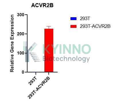
Figure 1. Characterization of ACVR2B over-expression in the 293T-ACVR2B stable clone using qPCR.

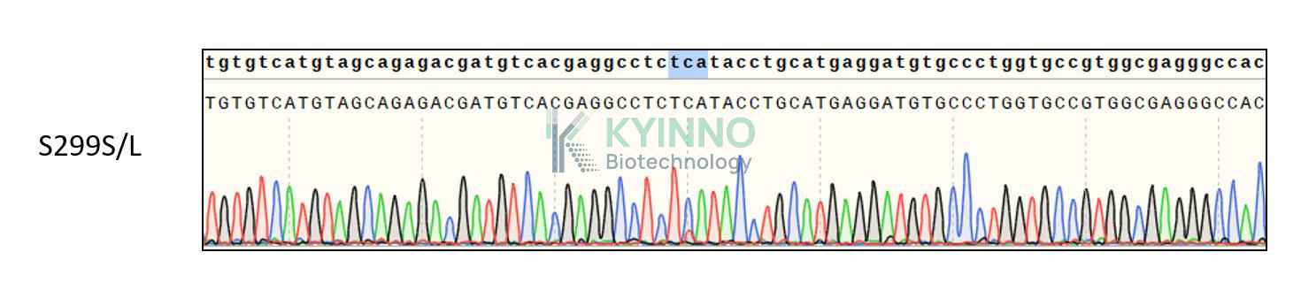
Figure 2. Characterization of ACVR2B over-expression in the 293T-ACVR2B stable clone using PCR.

Cell Resuscitation

  1. Pre-warm complete culture medium (basal medium and 10% FBS) in a 37°C water bath.
  2. Rapidly thaw the cryovial in a 37°C water bath for 1-2 minutes with gentle agitation.
  3. Transfer the vial to a biosafety cabinet, and disinfect the exterior with 70% ethanol.
  4. Aseptically transfer the cell suspension dropwise into a sterile centrifuge tube containing 9.0 mL of pre-warmed complete medium.
  5. Centrifuge at approximately 125 × g for 5–7 minutes at room temperature, carefully aspirate the supernatant without disturbing the cell pellet.
  6. Gently resuspend the pellet in an appropriate volume of complete medium and transfer the suspension into a T25 flask.
  7. Incubate the flask in a 37°C in a humidified 5% CO₂ incubator.
  8. Assess cell viability and morphology after 24 hours. If cells appear healthy, replace the medium with fresh medium supplemented with the appropriate selective antibiotic.
  9. Subculture the cells at a ratio of 1:4–1:6 every 2–3 days upon reaching 80%–90% confluency.

Cell Freezing

  1. Prepare the freezing medium (70% basal medium, 20% FBS and 10% DMSO) freshly before use.
  2. Pre-chill the freezing medium on ice and label the cryovials accordingly.
  3. Transfer the cell suspension to a sterile conical tube and perform a cell count to determine total viability and density.
  4. Centrifuge the cells at 250×g for 5 minutes at room temperature; carefully aspirate the supernatant.
  5. Gently resuspend the pellet in chilled freezing medium, ensuring a minimum cell density of 3×10⁶ cells/mL.
  6. Aliquot 1 mL of the cell suspension into each pre-labeled cryovial.
  7. Place the cryovials into a CoolCell® container and store at -80°C overnight for controlled-rate cooling.
  8. Transfer the cryovials to the liquid nitrogen for long-term storage the following day.

References

  1. Oprea S, Dumitru AV, Dumitrescu D et al. Mechanotransduction Failure and Molecular Rescue in Gastric Cancer: Kinetotherapy Across the IL-6/STAT3-Myostatin/ACVR2B-Akt/mTOR Axis. Medical sciences (Basel, Switzerland). 2026 Jul 1;14(3):. PMID: 42506334. doi: 10.3390/medsci14030365.
  2. Liu G, Liu Y, Zhao H et al. INHBB Promotes Colorectal Cancer Progression by Inducing CD8(+) T Cell Exhaustion via the ACVR2B-SMAD Signaling Axis. Cancer science. 2026 Sep 28;:. PMID: 42803598. doi: 10.1111/cas.70542.
  3. Patel JH, Tajer B, White J et al. Acvr2b receptors transduce all BMP signaling in the zebrafish gastrula and restrict Fibrodysplasia Ossificans Progressiva ACVR1-R206H signaling in a dose-dependent manner. bioRxiv : the preprint server for biology. 2025 Oct 7;:. PMID: 41279820. doi: 10.1101/2025.10.07.680911.

Use License Agreement

Research Use Only.
Not for use in diagnostic procedures or therapeutic applications.
Redistribution of the cell line or its derivatives is prohibited without prior written permission from Kyinno Biotechnology.